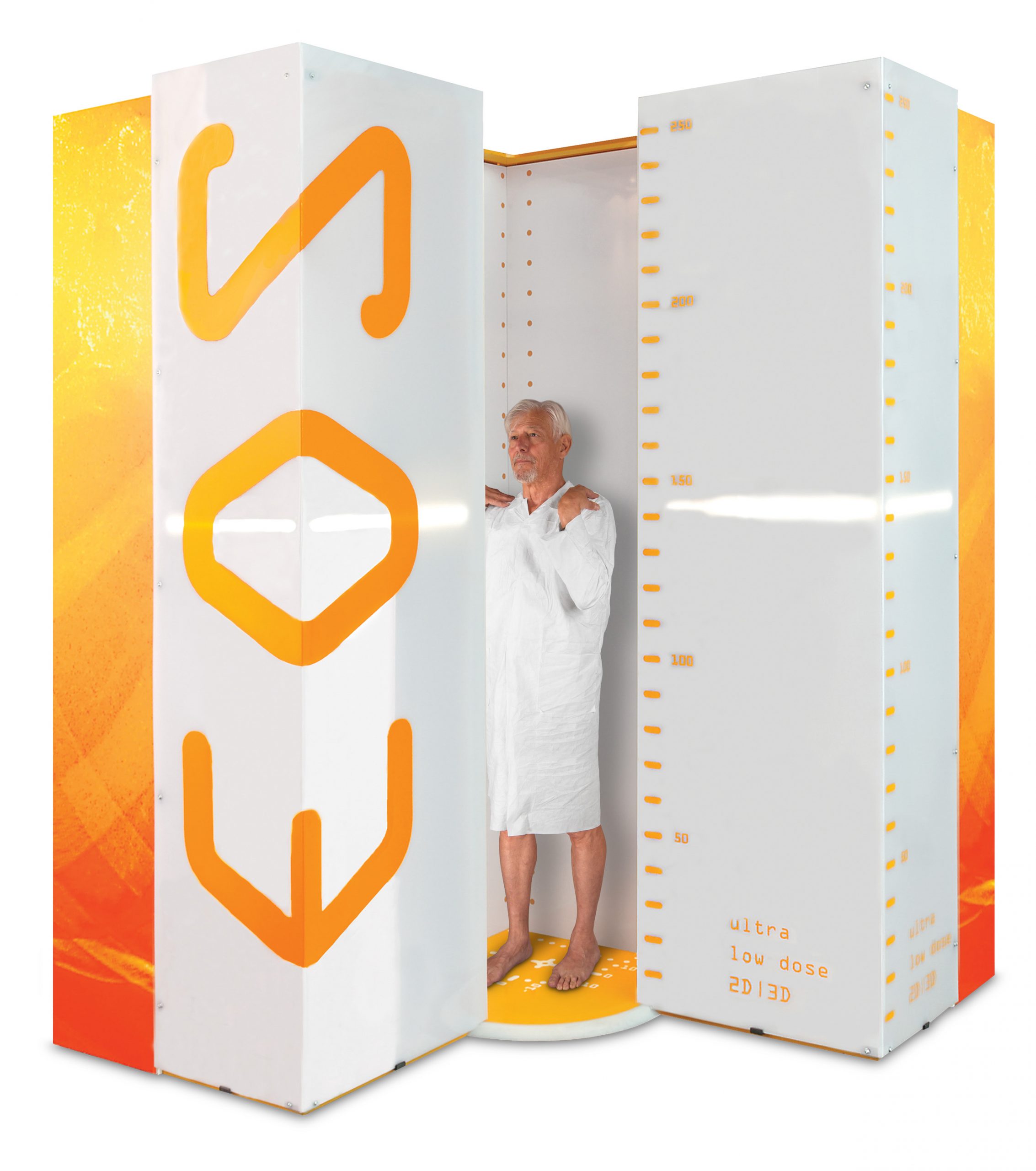
The EOS Imaging System is a low dose medical imaging tool that develops a full body 2D or 3D view of the spine, hip and knees. This low dose imaging offers patients a new standard of care with minimal exposure while collecting data all at once. EOS Imaging is a global medical device company who originates from French scientific research awarded the Nobel Prize in Physics in 1992.
What Makes the EOS Imaging System Beneficial?
Safe exams at the lowest radiation dose available vs the traditional X-ray exam. Exams of the patient full body at one visit and connecting the data from the imaging to 2D/3D workstation(sterEOS) and online 3D surgical planning options (EOSapps). Patients of all ages are looking for safer, better outcomes for their musculoskeletal conditions and the EOS Imaging system can provide this emitting 95% less radiation than a CT scan and up to 80% less radiation than the traditional X-ray, according to the National Library of Medicine.
Advanced Medical Care
Our bodies are 3D and weight bearing, therefore the EOS Imaging is completed while standing or sitting to provide the highly accurate 3D representations of your anatomy and pressure on your spine, which will then provide your healthcare provider with a seamless treatment plan using the EOS imaging platforms (EOSedge and EOS) with EOS Advanced Orthopedic Solutions.
Accuracy
A traditional X-ray provides one image on a film that would have to be pieced together, whereas the EOS imaging system has vertical traveling arms supporting X-ray beams. These X-ray beams acquire full body, weight bearing images that are then used to create the 3D model of the skeleton providing the most accurate image for your healthcare provider.
Convenience
EOS imaging provides reduced exam times, approximately 15-30 seconds for both children and adults, as well as reduced amounts of radiation for patients with instant results. For patients that need multiple images during their treatments and care, this reduced time and exposure is beneficial to their overall health.


